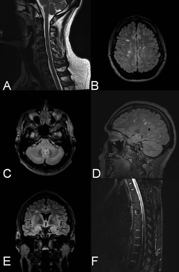
Figure 1. Patient’s MRI scans after eight months on ATRIPLA. A. Cervical MRI; Sagittal T2-weighted image reveals hypertensive lesions (C2-C3, C4, C5-C6). B-C. Axial FLAIR (Fluid-attenuated inversion recovery) brain images, with characteristic chronic periventricular and juxtacortical lesions (B), and a cerebellum lesion (C). D. Sagittal FLAIR brain image with hypertensive lesions involving the corpus callosum (Dawson fingers). E. Coronary FLAIR image with right juxtacortical hypertensive lesion. F. Sagittal T2-weighted thoracic MRI, with no apparent intramedullary demyelinating lesions, T5 spinal hemangioma.

