
Figure 15.
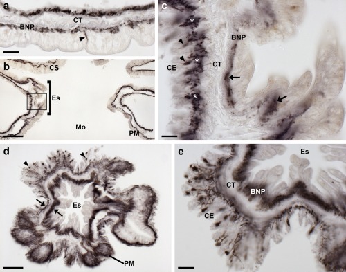
Localization of ArPPLN1b immunoreactivity in the peristomial membrane and esophagus of A. rubens. (a) An immunostained bipolar shaped cell can be seen here in the external epithelial layer of the peristomial membrane (arrowhead) and in processes located in the underlying basiepithelial nerve plexus. Immunostaining is also present in cells and processes located beneath the coelomic epithelium, which is separated from the basiepithelial nerve plexus by a layer of collagenous tissue. (b) Transverse section of the central disk showing immunostaining in the peristomial membrane, the esophagus and the cardiac stomach. The boxed region is shown at higher magnification in panel (c). (c) Longitudinal section of the esophagus (in a transverse section of the central disk) showing immunostained cells (arrowheads) and processes (asterisks) beneath the coelomic epithelium and stained processes (arrows) in the basiepithelial nerve plexus beneath the epithelial layer that forms the external lining of the esophagus. (d) Horizontal section of the central disk at the level of the junction between the esophagus and the peristomial membrane showing immunostained cells (arrowheads) and processes (arrows). (e) High magnification transverse section of the esophagus (in a horizontal section of the central disk) showing immunostained cells and processes beneath the folded coelomic epithelium and dense immunostaining in the basiepithelial nerve plexus beneath the epithelial lining of the eosophagus lumen. Abbreviations: BNP, basiepithelial nerve plexus; CE, coelomic epithelium; CS, cardiac stomach; CT, collagenous tissue; Es, esophagus; Mo, mouth; PM, peristomial membrane. Scale bars: 20 μm in (a), (c), (e); 100 μm in (b); 50 μm in (d)