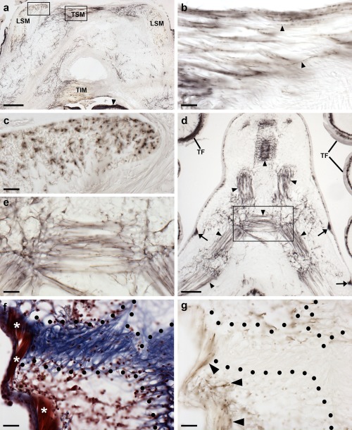
Figure 18.

Localization of ArPPLN1b immunoreactivity in the innervation of interossicular muscles in A. rubens. (a) Transverse section of the ambulacrum showing immunostaining associated with muscles linking the ambulacral ossicles, which include the TSM, TIM, and LSM. The intensely stained radial nerve cord (arrowhead) can also be seen in this image. The boxed regions are shown at higher magnification in panels (b) and (c). (b) Immunostained nerve fibers (arrowheads) in a longitudinal section through the TSM. (c) Immunostained profiles of nerve fibers in a transverse section through the LSM. (d, e) Immunostaining in the body wall at the junction between two arms in a juvenile starfish. Immunostained fibers can be seen associated with muscles that link adambulacral ossicles (arrowheads). Stained fibers are also evident in thickenings of the sub‐epithelial nerve plexus of the body wall (arrows) and in the tube feet. A high magnification image of the boxed region is shown in (e). (f) Trichrome stained section of the body wall showing an interossicular muscle (white asterisks) and collagenous tissue (area bounded by black dots) linking adjacent ossicles. (g) Immunostained section adjacent to the section shown in panel (f). By comparing the immunostaining with the trichrome staining it can be seen that the immunostained fibers are associated with the interossicular muscle but not with the collagenous tissue (area bounded by black dots). Abbreviations: LSM, longitudinal supra‐ambulacral muscle; TF, tube foot; TIM, transverse infra‐ambulacral muscle; TSM, transverse supra‐ambulacral muscle. Scale bars: 200 μm in (a); 20 μm in (b), (c), (e), (f), (g); 100 μm in (d)
