
Figure 2.
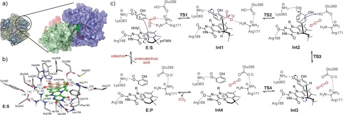
AroY structure and suggested mechanism based on calculations. a) Hexameric quaternary structure of EcAroY. Dimer pairs are shown in ribbon representation in green and blue. The cryo‐EM envelope is shown as a gray translucent surface. A detailed view of the EcAroY monomeric structure (in the circle) showing the prFMN‐binding domain (blue), oligomerization domain (green), and C‐terminal helix (red) is also given. b) Optimized structure of the active‐site model employed in the computational study. Atoms marked with asterisks were fixed during the geometry optimization. The prFMN cofactor is shown in green and the substrate in salmon ball‐and‐stick presentation. Distances are given in Å. For clarity, only polar hydrogen atoms and the hydrogen atoms on the substrate are shown. c) Reaction mechanism suggested on the basis of the calculations.