Figure 1.

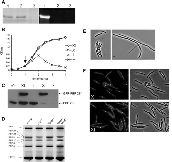
A. Penicillin binding activity of wild type and mutant forms of PBP 2B. PBP 2B and PBP 2B(S309A) were overproduced in E. coli and the membrane fraction was purified, labelled with Bocillin‐FL and separated by SDS‐PAGE. The left panel is an image of the protein gel following Coomassie staining showing that similar amounts of total protein and overproduced PBP 2B protein were present on the gel and the right panel shows an image of the same gel when scanned for fluorescence. The gel was loaded with membrane preparations from E. coli over expressing WT pbpB (lane 1); pbpB* (2); control (vector only) (3).
B. Growth curve of strain 4004 in various inducer conditions. Strain 4004, which contains the pbpB* allele controlled by a xylose induced promoter (Pxyl) and a WT pbpB controlled by an IPTG induced promoter (Pspac), was grown in PAB with IPTG then transferred to Fresh PAB media with various combinations of supplements (XI, media supplemented with xylose and IPTG; X, xylose alone; I, IPTG alone; ‐, no addition.) and the growth against time was measured by optical density.
C. Inducer dependence of PBP 2B and GFP‐PBP 2B. Western blot of total protein samples taken at the start of the experiment (t0) and 1 h after removing or adding inducers (arrow in panel B) and probed with polyclonal antisera specific for PBP 2B.
D. An image obtained by fluorography showing the binding profile of bocillin–FL to B. subtilis PBPs from strains 168 (wild‐type), pbpB*, ΔpbpH and ΔpbpH pbpB*. The identity of each PBP is indicated on the left of the image.
E. Phase contrast images of strain 4004 grown in the presence of IPTG (I) or in the absence of any inducers (‐).
F. Localisation of GFP‐PBP 2B(S309A) in cells in the presence or absence of WT pbpB expression. Cells of strain 4004 grown with xylose and IPTG (XI) and xylose alone (X). Each panel shows a fluorescence micrograph and the corresponding phase contrast image.
