Figure 2.

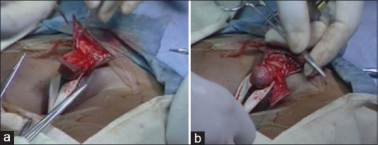
(a) Dartos flap raised by dissecting between the skin and dartos layer of dorsal prepuce. (b) Dartos fascia brought ventrally and then de-epithelialized to cover the ventral suture line

(a) Dartos flap raised by dissecting between the skin and dartos layer of dorsal prepuce. (b) Dartos fascia brought ventrally and then de-epithelialized to cover the ventral suture line