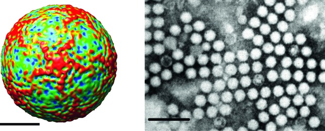
Fig. 1.
(Left) Surface view of the virion of infectious flacherie virus along a five-fold axis reconstructed by cryo-electron microscopy. The bar represents 10 nm (courtesy of J. Hong). (Right) Negative contrast electron micrograph of the isometric particles of an isolate of infectious flacherie virus. The bar represents 100 nm (courtesy of H. Bando).

