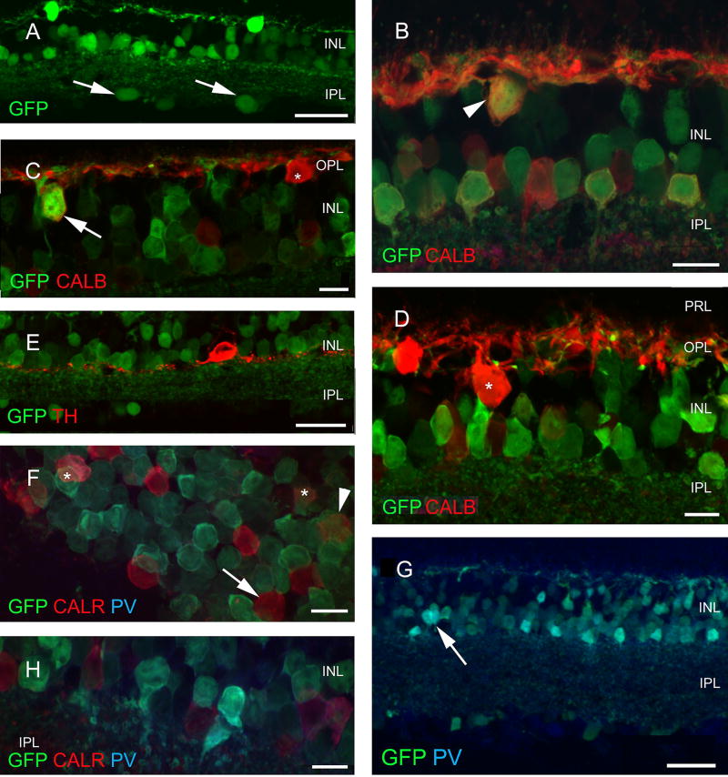
Fig. 4.
Retinal expression pattern of GAD65-GFP construct. Several cell types show GFP expression in the GAD65-GFP construct, including large cells in the ganglion cell layer (arrows in a). Among interneurons, some (but not all) horizontal [arrowhead weak label in b, arrow strong positivity in c and none (asterisk) in d] cells express GFP. Among the amacrines, TH- (e) and CALB-containing cells (b) do not, while CALR-containing cells (arrowhead and asterisks in f) rarely contain GFP; no colocalization is seen in g (arrow) while small, narrow-field PV-positive cells (arrow) frequently express GFP (g, h). Scale bars 30 µm in a, e and h and 10 µm in all other figures. PRL photoreceptor layer, ONL outer nuclear layer, OPL outer plexiform layer, INL inner nuclear layer, IPL inner plexiform layer, GCL ganglion cell layer, GFP green fluorescent protein, PNA peanut agglutinin, TH tyrosine hydroxylase, CALR calretinin, CALB calbindin 28 kDa, PV parvalbumin

