Fig. 6.
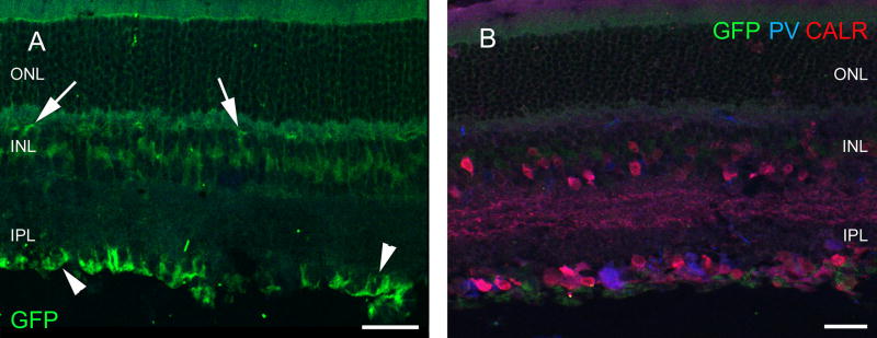
Retina of the PVRosa-YFP strain. GFP is present in the glial cells only (arrows), labeling is strong in the endfeet of glial cells (arrowheads) in the inner limiting membrane (a). At the same time, cells can be labeled for PV and CALR in the retina of this strain as usual (b). Scale bars 40 µm in a and 30 µm in b. PRL photoreceptor layer, ONL outer nuclear layer, OPL outer plexiform layer, INL inner nuclear layer, IPL inner plexiform layer, GCL ganglion cell layer, GFP green fluorescent protein, PNA peanut agglutinin, TH tyrosine hydroxylase, CALR calretinin, CALB calbindin 28 kDa, PV parvalbumin

