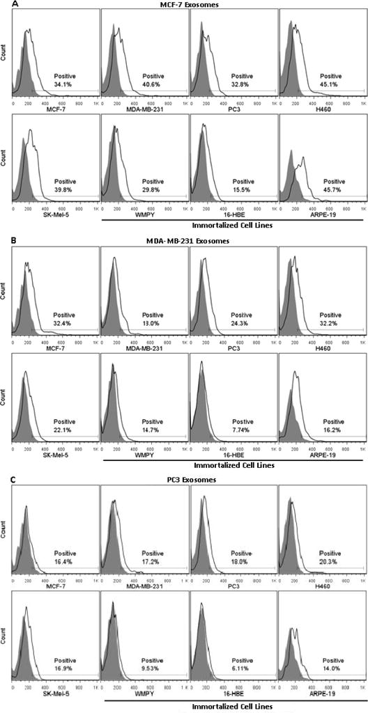
Figure 4.

Exosome association with cancer and immortalized cell lines. Exosomes isolated from the supernatant of MCF-7, MDA-MB-231, and PC3 cell lines were added back to the media of five cancer cell lines (MCF-7, MDA- MB-231, PC3, H460 and SK-Mel-5) and three immortalized cell lines (WMPY, 16-HBE and ARPE-19). Exosomes were labeled with 0.003% DID by weight; the greater association of exosomes as compared to liposomes required lower levels of DID to avoid detector saturation. Extent of association was measured using flow cytometry and quantified by monitoring the percentage of cells that had increased fluorescence when compared to control cells (“Positive”). Results from a single experiment are presented; repeat experiments yielded virtually identical results.
