
Figure 7.
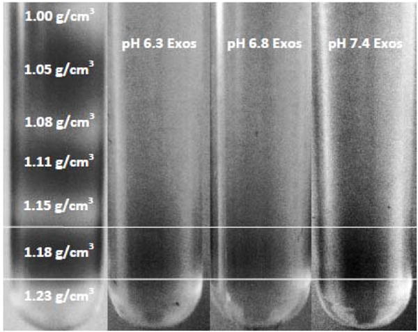
Density of isolated exosomes. Exosomes labeled with 0.01% DID in PBS loaded onto a continuous sucrose gradient and centrifuged at 200k × g for 10 hrs. The fluorescent dye, DID, allowed for visual identification of exosomes. In order to distinguish between each sucrose layer, a control sucrose gradient was run (far left) where every other sucrose layer was stained with fluorescein. Exosomes isolated from the supernatant of PC3 cells cultured at pH 6.3, 6.8 and 7.4 all partitioned into the sucrose layer corresponding to a density of 1.18 g/cm3.