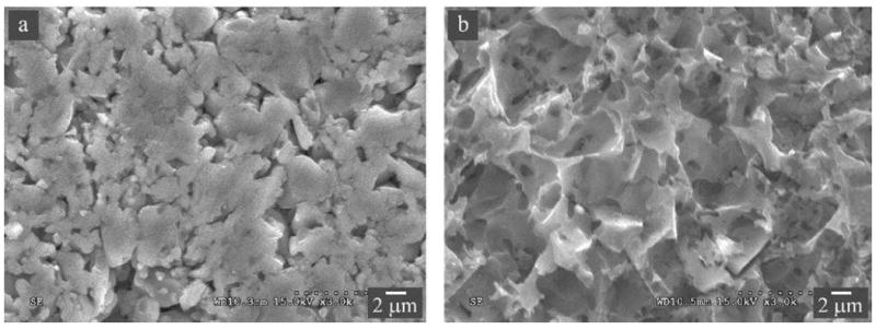
Figure 5.

Microstructure of Vita Enamic observed using secondary electrons in a SEM. (A) A polished and then thermally etched surface, revealing a ceramic network structure consisting of ~25 vol% porosity following selective removal of the polymer phase. (B) A polished and then acid etched surface, showing the polymer network after selective removal of the surface ceramic material.
