
Figure 7. In ACE 3/9 mice, the only source of renal ACE are the tubular epithelial cells.
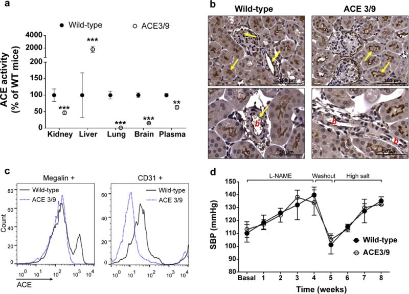
ACE activity was assessed in different tissues. (a) Values represent mean ± SEM; n = 5 per group, **P < 0.01 and ***P < 0.001 versus wild-type mice. Renal ACE expression was evaluated by (b) immunohistochemistry, where b indicates blood vessel, and by (c) flow cytometry. Megalin and CD31 were used as markers of proximal tubular cells and endothelial cells, respectively. (d) Wild-type and ACE 3/9 mice were exposed to the post–L-NAME protocol (n = 5).