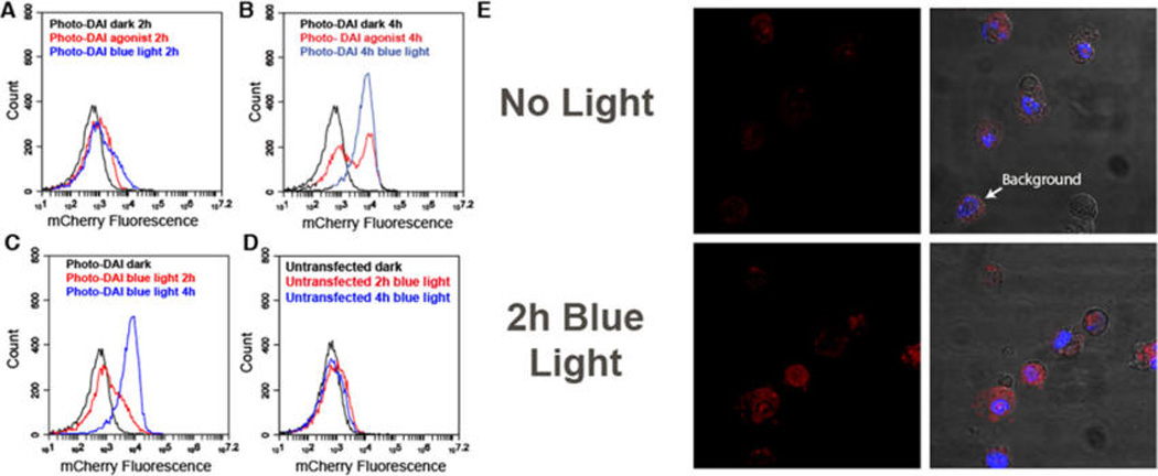
Figure 3.

Fluorescent reporting of dimerization from cells expressing photo-DAI. (A) Flow cytometry of HEK cells expressing photo-DAI after 2 h of exposure to either blue light or the native DAI agonist (100 ng/mL Poly(dA:dT)). Cells incubated for 2 h in the dark (black), with Poly(dA:dT) (red), or with blue light (blue). (B) Cells expressing photo-DAI exposed to either blue light or Poly(dA:dT) for 4 h. Cells incubated for 4 h in the dark (black), with Poly(dA:dT) (red), or with blue light (blue). (C) Cells transfected with photo-DAI exposed to blue light for 0 h, 2 h, or 4 h. Cells incubated in the dark 4 h (black) and cells exposed to 2 h blue light (red) or 4 h (blue). (D) Untransfected cells exposed to blue light for 0 h, 2 h, or 4 h. Cells incubated in the dark 4 h (black) and cells exposed to blue light 2 h (red) or 4 h (blue). (E) Microscope images of HEK cells expressing photo-DAI Left: mCherry channel. Right: merge channel of mCherry and DAPI. Top: incubated in the dark. Bottom: 2 h blue light exposure.
