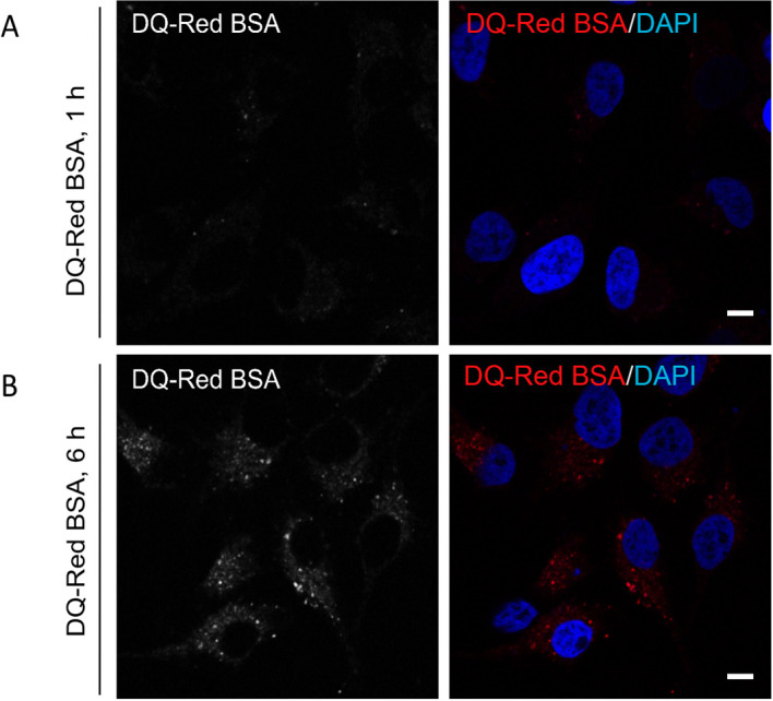
Figure 2. De-quenching of DQ-Red BSA fluorescence at different time points post endocytosis.

Representative single-plane confocal micrographs of HeLa cells showing fluorescence (red signal) of DQ-Red BSA after an uptake of 1 h (A) and 6 h (B) time points. At the end of the uptake time point, cells were fixed and stained with DAPI (blue) to mark the cell nucleus. A bright fluorescence punctae of DQ-Red BSA is visible post 6 h uptake as compared to 1 h, indicating its delivery to acidic compartments of the cell. Scale bars = 10 µm.
