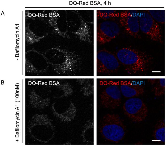
Figure 3. DQ-BSA uptake and delivery to lysosomes.

Representative single-plane confocal micrographs of HeLa cells incubated with DQ-Red BSA in 1% serum containing media for indicated time point in the absence (A) or presence (B) of 100 nM Bafilomycin A1, a V-ATPase inhibitor that disrupts lysosome fusion. The cell nucleus is stained using DAPI (blue). Scale bars = 10 µm.
