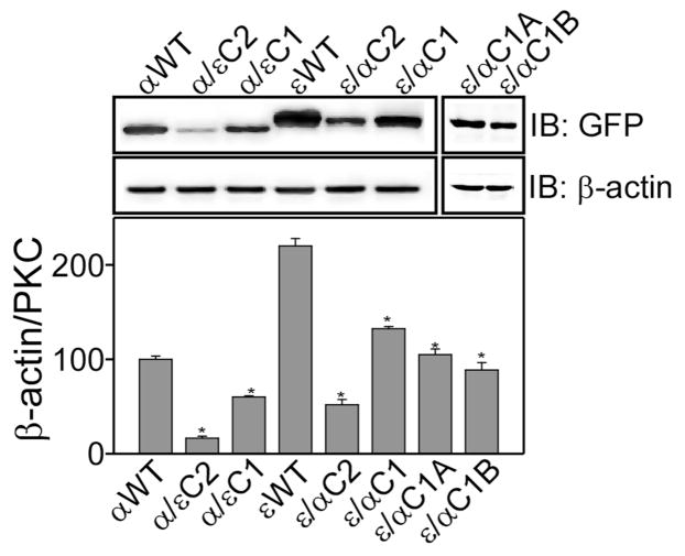
Figure 2.
Effect of domain swapping on the protein expression in HEK293 cells. Upper panel shows Western blot analysis of wild type and mutant proteins in HEK293 cells. Cells were transiently transfected with the particular plasmid, cells were lysed after 48 h and whole cell lysate (40 μg) were used for immuno-blotting. Expressed proteins were detected using anti-GFP antibody and anti-β-actin antibody was used as a loading control. The lower panel shows bar graph of densitometry analysis of the upper panel. Results are displayed as Mean ± SEM, n = 3 and one-way ANOVA (Bonferroni post hoc test) was used for statistical significance analysis. *, P values less than 0.05 were considered significant.

