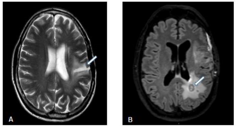
Figure 1.
(A) Axial T2 weighted axial MRI image of leukoencephalopathy (arrow) occurring in a 53 year old female patient 5 years after receiving chemoradiation (60 Gy) treatment for a high grade glioma. (B) Axial FLAIR MRI image of radionecrosis as a hypoenhancing center (arrow) surrounded by a rim of enhancement indicating active inflammation in a 48 year old male patient following chemoradiation (60 Gy) treatment for high grade glioma.

