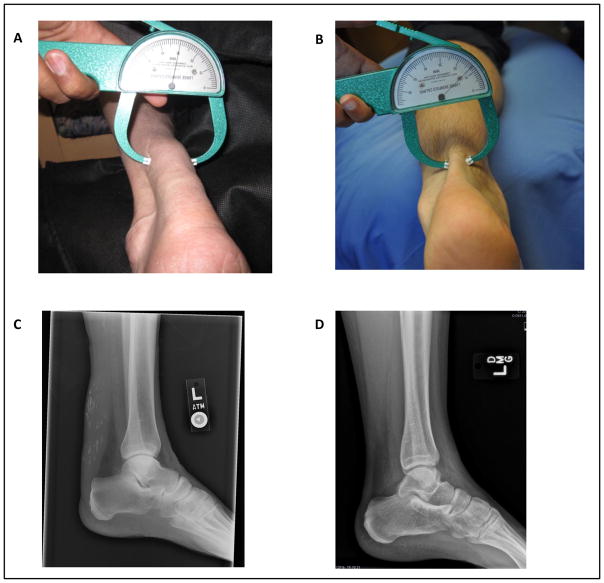
Figure 1.
Achilles tendon xanthomas. A, Image of patient’s Achilles tendon with caliper measuring thickness of 30 mm. B, Image of a normal Achilles tendon with width of 13 mm. C, Lateral plain radiograph demonstrating thickness of the patient’s Achilles tendon with scattered calcifications. D, Lateral plain radiograph with normal Achilles tendon.

