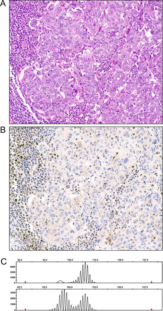
Figure 1.

MSI-high endometrial carcinoma with PMS2 loss by immunohistochemistry. (A) H&E of tumor, demonstrating a poorly differentiated endometrial carcinoma with adjacent tumor infiltrating lymphocytes. (B) PMS2 immunohistochemistry, showing that tumor infiltrating lymphocytes and stromal cells adjacent to the tumor have retained nuclear expression for PMS2. Tumor cells have faint cytoplasmic expression of PMS2, but no nuclear expression for this mismatch repair protein. (C) A representative microsatellite tracing for BAT40 from this MSI analysis. Note that tumor tracing has more peaks than the tracing for normal. In this case, normal ovary was used as a source of normal. This tumor had allelic shift in 4 other microsatellites as well (not shown).
