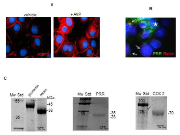
FIGURE 1.
(A) Characterization of M-1 collecting duct cell line. M-1 cells can increase the abundance of aquaporin 2 (AQP-2, red) after vasopressin (AVP) treatment. (B) Co-labeling of (pro)renin receptor (PRR, green) and prorenin-renin (red) showing differential distribution, probably in intercalated cells (asterisks) and principal cells (arrows) respectively. (C) Recombinant proteins used as positive controls to establish antibody specificity and migration.

