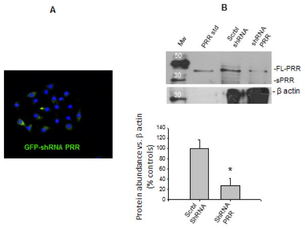
FIGURE 3.
(A) Green fluorescent protein (GFP) positive cells confirming effectiveness of GFP-shRNA-PRR transfections, which reached 70 ± 8% (n = 10). (B). Effectiveness of PRR silencing verified by band intensity compared with non-transfected cells and cells transfected with scramble-shRNA (73 ± 14% P < 0.05 vs. scramble-shRNA transfected group, n = 6). Western blot membrane was revealed by quimioluminescent method after incubation with rabbit anti-ATP6AP2 and mouse anti-β-actin. A positive control (recombinant PRR) is also shown. Mw: Molecular weight standard.

