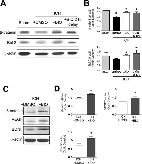
Figure 2.

Regulatory effects of BIO on β-catenin expression and other factors in the ICH brain. (A) Representative Western blot images of perihematoma tissue lysates collected at 2 days post-ICH. (B) Quantification of immunoblots of β-catenin and the antiapoptotic factor B-cell lymphoma 2 (Bcl-2). Samples are normalized to β-actin loading control, and groups are normalized to sham. (C) Representative Western blot data of perihematoma tissue lysates at 14 days post-ICH to evaluate long-term BIO effects. (D) Quantification of immunoblot intensities of β-catenin, vascular endothelial growth factor (VEGF), and brain-derived neurotrophic factor (BDNF). Samples are normalized to β-actin loading control, and groups are normalized to ICH + dimethyl sulfoxide (DMSO) group. Mean+SEM. ∗p < 0.05 compared to ICH + DMSO group. #p < 0.05 compared to sham. n = 3 mice for sham group and n = 6–8 for other groups.
