
Figure 3.
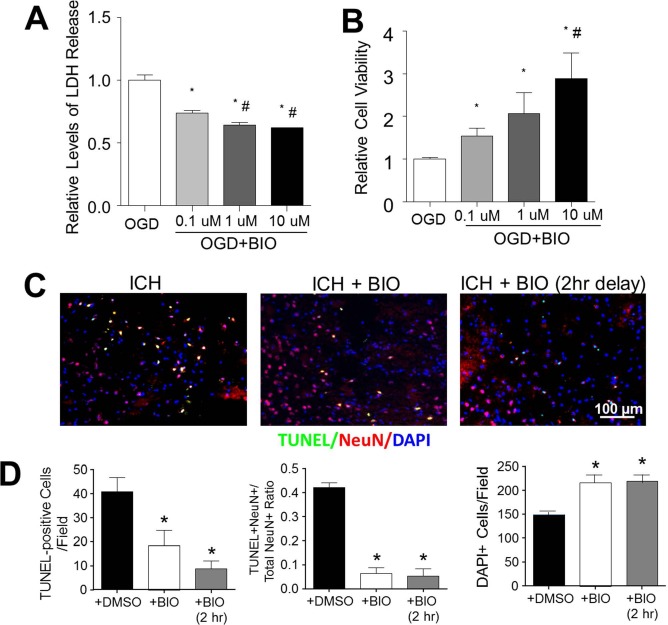
BIO-enhanced viability of neurons in vitro and in the ICH brain. (A) Lactate dehydrogenase (LDH) release correlated with neuronal cell death following oxygen-glucose deprivation (OGD). A dose-dependent neuroprotective effect was observed in BIO-treated cultures. (B) Relative cell viabilities determined by the 3-(4,5-dimethyl-thiazol-2-yl)-2,5-diphenyltetrazolium (MTT) assay in OGD with and without BIO treatments. Mean+SEM. ∗p < 0.05 compared to OGD + DMSO group. #p < 0.05 compared to OGD + 0.1 μM BIO group. (C) Representative images of multiplexed immunofluorescence of terminal deoxynucleotidyl transferase 2′-deoxyuridine 5′-triphosphate (dUTP) nick-end labeling (TUNEL, green), neuronal nuclei (NeuN, red), and 4′,6-diamidino-2-pheylindole (DAPI, blue). (D) Quantification of stereological counts of total cell numbers (DAPI+), numbers of cell death (TUNEL+ cells), and numbers of neuronal cell death (TUNEL+/NeuN+ colabeled cells, yellow signal). Mean + SEM. ∗p<0.05 compared to ICH + DMSO group. n = 4–5 culture batches/group.