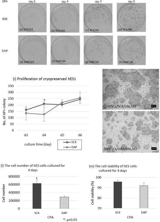
Figure 4.

Proliferation profiles of human embryonic stem cells (hESCs) cryopreserved with SCK or a solution of 2 M dimethyl sulfoxide, 1 M acetamide, and 3 M DAP using AP staining. AP-stained hESCs cryopreserved with SCK (a-d) or DAP (e-h) after 3, 4, 5, and 6 days of culturing (day 3, day 4, day 5, and day 6). (i) Comparison of the number of AP+ hESC colonies after preservation with SCK or DAP and culturing for 3, 4, 5, and 6 days. AP+ colonies were counted in four fields at low magnification (10x). hESC colonies 3 days after cryopreservation in SCK (j) or DAP (k). AP staining was observed under an inverted microscopy at 40x magnification. The number (l) and viability (m) of hESCs cultured for 4 days after cryopreservation in SCK or DAP. All data are presented as means ± SD. Statistical evaluations were done by Student's t-test, *p < 0.05, statistically significant difference. Scale bars: 100 um. CPA, cryoprotective agent.
