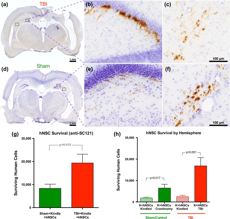
Figure 2.
Long-term survival (14 wpt) of hNSCs is significantly greater in TBI animals than in shams; survival was also better in the nonkindled hemisphere. At 14 wpt and 1 wk after stimulating to measure AD, transplanted hNSCs show long-term survival in the brains of both sham and TBI ATN rats. (a–c) Immunohistochemistry of transplanted hNSCs with the human cytoplasmic protein marker, anti-SC121, showing survival and engraftment in both injured (a) and sham (d) brains in photomontage collected at 2.5×. Boxed areas in part figures a and d shown at higher magnification in b, c e, and f, respectively, taken at 20×. (g) Stereological quantification of transplanted hNSCs in sham (n = 13, 8,337 ± 1,812) and in TBI animals (n = 14, 19,344 ± 3,816) shows a significant difference in cell survival between experimental groups (2-tailed t test t25 = 2.54, p = 0.0177). (h) Stereological quantification of transplanted hNSCs shows a significant difference between the kindled side and the contralateral side in both groups (Sham—2-tailed t test t24 = 2.56, p = 0.017; TBI—2-tailed t test t26 = 3.65 p = 0.001). Data are presented as mean ± standard error of the mean (SEM), with TBI animals demonstrating significantly higher survival. Animals were kindled on the contralateral side of the injury (CCI injured for TBI animals and craniotomy for sham). wpt,: weeks posttransplantation; hNSC, human neural stem cell; TBI, traumatic brain injury; AD, after discharge; ATN, immunodeficient athymic nude; anti-SC121, human cytoplasmic marker; CCI, controlled cortical impact; K, kindled.

