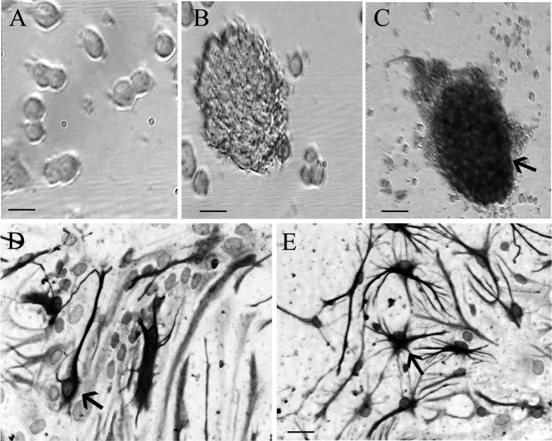
Figure 1.
Morphology and identification of neural stem cells (NSCs). (a) The morphology of cells at 12 h of primary culture. (b) The neurospheres composed of hundreds of cells at 3 d after culture under microscope in bright field. (c, d, and e) Three days after culture, respectively. Nestin (marker of NSCs), NeuN (marker of neurons), and glial fibrillary acidic protein (GFAP; marker of astrocytes) immunopositive cells were observed under the light microscope, respectively. Scale bar = 25 μm in a, b, d, e. Scale bar = 50 μm in c.

