
Figure 2.
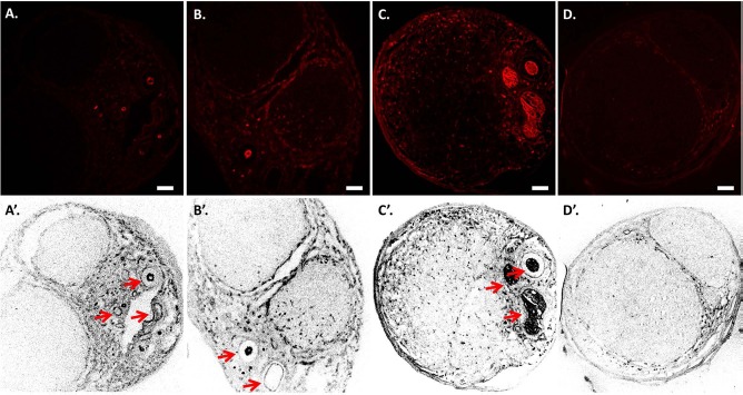
Revascularization of the decellularized allogenic nerve matrix (DANM). Immunohistochemistry for mouse monoclonal anti-rat endothelial cell cytoplasmic antigen (RECA-1) was performed on the transverse sections obtained from the midportion of the tube in the VD group at 5 days (A, A'), at 1 week (B, B'), and at 4 weeks (C, C') and in the D group at 4 weeks (D, D'). (A, A') A few RECA-1+ cells were detected only at the periphery of the DANM, and no RECA-1+ cells were observed inside the DANM at 5 days. (B, B') RECA-1+ cells were observed in the DANM at 1 week. The revascularized area was located mainly beside the inserted sural vessels. (C, C', D, and D') At 4 weeks, the RECA-1+ cells had spread diffusely in the DANM in the VD group (C, C'), whereas RECA-1+ cells were not found in the DANM in the D group (D, D'). The arrows indicate the inserted vascular bundle. Scale bars: 100 μm. VD group, the group with implantation of the sural vessels and a decellularized allogenic nerve matrix.