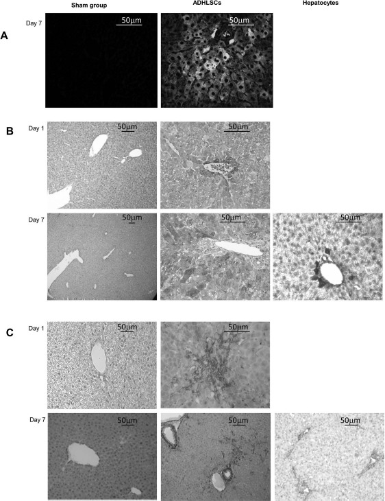
Figure 5.

Visualization of the human cells implanted in mouse livers by immunohistochemistry. (A) Representative result of the tracking of eGFP+ ADHLSCs in mouse liver tissue by direct fluorescence microscopy. Implantation of human cells in group B is visible 7 days after injection, with cells primarily located around the periportal spaces and starting to enter the parenchyma. (B) Representative result of the anti-human albumin immunostaining. Liver sections from mice transplanted with ADHLSCs and the sham group were analyzed on days 1 and 7 following transplantation (only on day 7 for hepatocytes). One day after transplantation, human albuminpositive ADHLSCs were only found implanted near the periportal spaces. Seven days after injection, human cells had migrated in the liver parenchyma. (C) Representative result of the anti-human α-smooth muscle actin (α-SMA) immunostaining. Liver sections from mice transplanted with ADHLSCs and the sham group were analyzed on days 1 and 7 following transplantation (only on day 7 for hepatocytes). α-SMA+ cells were detected in the mice injected with ADHLSCs, confirming the results obtained with the antihuman albumin staining, whereas no α-SMA+ cells were detected in the mice injected with hepatocytes, since these cells are α-SMA−. ADHLSCs, adult-derived human liver stem/progenitor cells.
