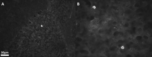
Figure 4.

Photomicrographs of the brain of a 58-day-old sHW rat 18 days after carotid injection of hNPCs. (A) A single hNPC detected in the cerebellum by immunofluorescent staining with anti-human nuclei antibodies. (B) Two hNPCs in the temporal lobe of the brain. Magnifications for both images were taken at 400×. sHW, spastic Han–Wistar rats; hNPCs, human-derived neural progenitor cells.
