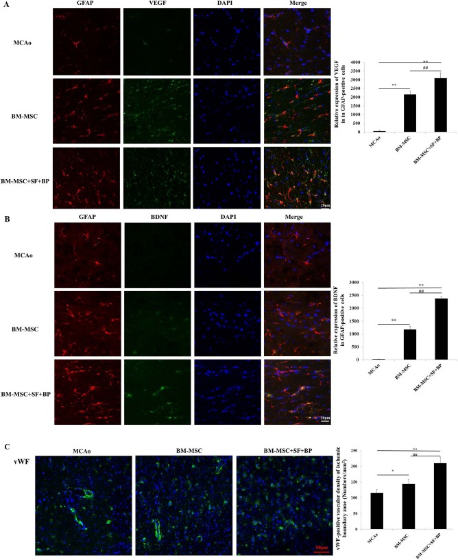
Figure 3.

Astrocyte-derived VEGF and BDNF expression and vWF+ vascular density in the ischemic boundary zone on the seventh day after stroke. The fresh-frozen coronal sections in each group were collected, and triple fluorescence immunostaining was performed. (A, B) Immunofluorescence stainings of GFAP (red), VEGF (green), and BDNF (green) were presented, and relative expressions of VEGF and BDNF in the GFAP+ area were analyzed. It indicated that combination of BM-MSCs with SF and BP notably improved astrocytic VEGF and BDNF expression. (C) vWF immunofluorescence staining (green) was presented, and positive signal was analyzed. Data are expressed as mean±SD. ∗p < 0.05, ∗∗p < 0.01, compared with MCAo; ##p < 0.01, compared with BM-MSC. The experiment was repeated three times, and representative pictures are shown. Scale bars: 20 μm (A, B), 50 μm (C). vWF, von Willebrand factor primary antibody; VEGF, vascular endothelial growth factor; BDNF, brain-derived neurotrophic factor; GFAP, glial fibrillary acidic protein; BM-MSCs, bone marrow-derived mesenchymal stem/stromal cells; MCAo, middle cerebral artery occlusion; SF, sodium ferulate; BP, n-butylidenephthalide; DAPI, 4′,6-diamidino-2-phenylindole.
