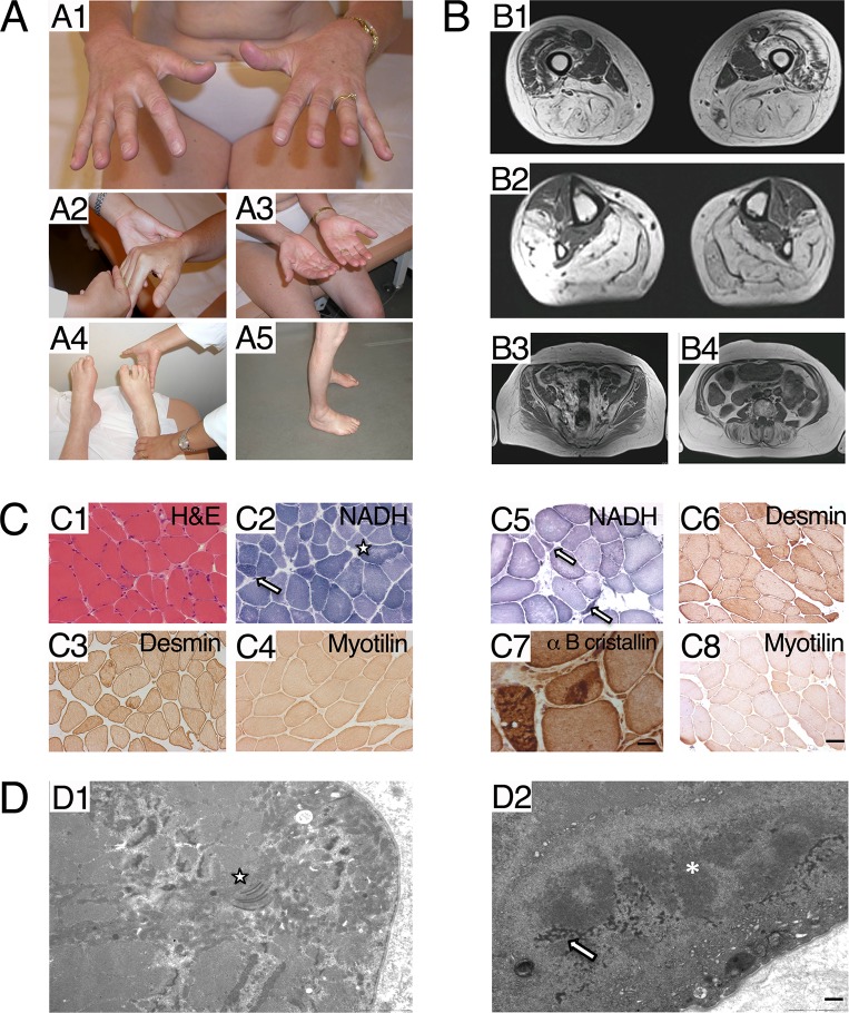
Fig 1. Clinical data.
A. Clinical data of the proband (III:3). A1, A2: Hand extensors weakness. A3: Palmar atrophy, mainly in thenar muscles. A4: Peroneus lateralis, markedly affected, as all legs muscles except for tibialis anterior and posterior muscles. A5: Leg atrophy. B. MRI of the proband. B1: Thigh. Hamstrings and adductors severely involved; quadriceps partially spared, except right vastus lateralis and intermedialis. B2: Legs. Peroneal lateralis severely affected, as all muscles except tibialis anterior and tibialis posterior, B3: Pelvic muscles spared in comparison with femoro-distal muscles. C. Morphological studies. C1-C4 Radialis muscle biopsy of proband (III:3). C1. Haematoxylin & Eosin staining: presence of some nuclear internalization and fiber size variation. C2 NADH: lobulated fibers are indicated by an arrow; uneven staining of the intermyofibrillar network in some fibers is indicated by a star. C3 desmin: mild diffuse desmin surcharge in several fibers C4 myotilin: normal staining. C5-C8 Peroneal muscle biopsy from the proband’s son (IV:2). C5 NADH: atrophic lobulated fibers are indicated by an arrow. C6 desmin: mild diffuse desmin surcharge in several fibers. C7 alpha B cristallin: presence of dense protein aggregates. C4 myotilin: normal staining (Scale bar = 20 μm for all images and 8 μm only in C7). D. Ultrastructural studies: Radialis muscle biopsy of proband (III:3), EM analysis. D1: presence of numerous abnormal mitochondria harbouring dotty or paracrystallin inclusions, in proximity of the star. D2: presence of a cytoplasmic protein aggregate composed by dark osmiophilic granulo-filamentous material corresponding to desmin, indicated by an arrow, and filamentous material indicated by an asterisk (Scale bar = 1 μm).

