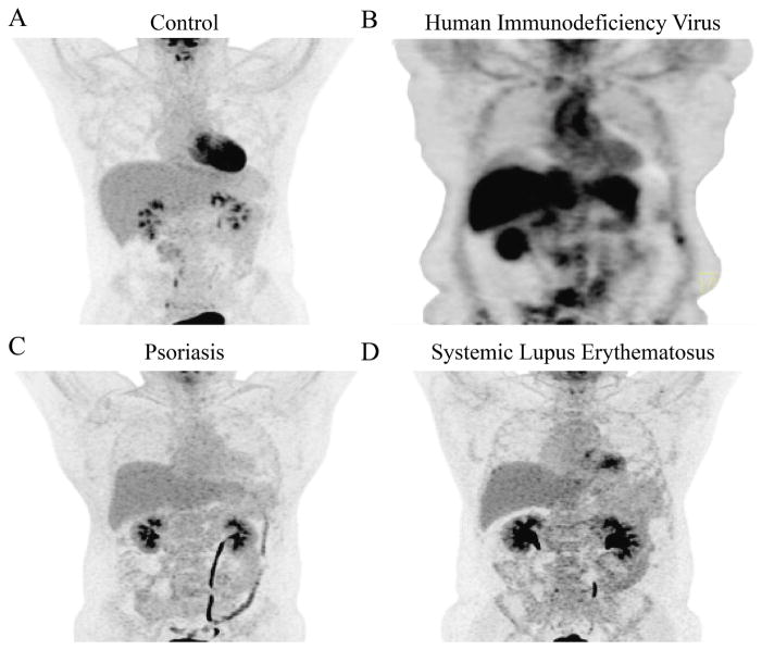
Figure 2. Vascular Images of Chronically Inflamed Human Models.
Representative 18F-FDG-PET/CT imaging of the aorta in a healthy volunteer (A), compared with the aortas of patients with human immunodeficiency virus (B), psoriasis (C), and systemic lupus erythematosus (D). CT = computed tomography; FDG = fluorodeoxyglucose; PET = positron emission tomography.

