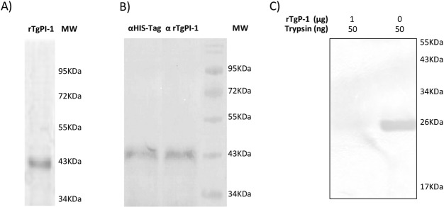
Fig 2. Purification and activity of rTgPI-1.
(A) SDS-PAGE of rTgPI-1 stained with Coomassie Blue. (B) Western blot of rTgPI-1 revealed with anti-His-Tag (lane 1) antibody or mouse anti-TgPI-1 serum (lane 2). (C) Analysis of inhibitory effect of rTgPI-1 on trypsin by gelatin substrate-SDS PAGE. All lanes contain 50 ng of trypsin. Lane 1, 1 μg of rTgPI-1; lane 2, control without rTgPI-1. Gelatinolytic activity was visualized by staining with Coomassie Brilliant Blue. The image was digitally inverted.

