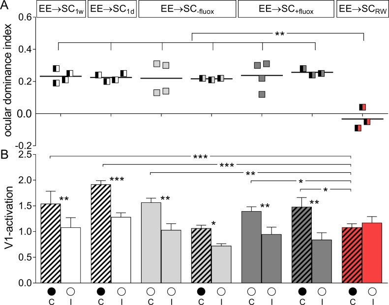
Fig 7. ODIs and V1-activation of EE-mice transferred to standard cages (SCs).
A. Optically imaged ODIs without and with MD of EE-mice transferred to SC for one week (EE→SC1w) or one day (EE→SC1d) before MD (white), EE→SC-fluox (light grey), EE→SC+fluox (dark grey) and EE→SCRW mice (red). B. V1-activation elicited by stimulation of the contralateral (C) or ipsilateral (I) eye without and after MD. Only in EE→SCRW mice showed an OD-shift after MD: both eyes activated V1 about equally strong whereas in all other groups, V1 continued to be dominated by the previously deprived contralateral eye. Layout and data display as in Fig 3.

