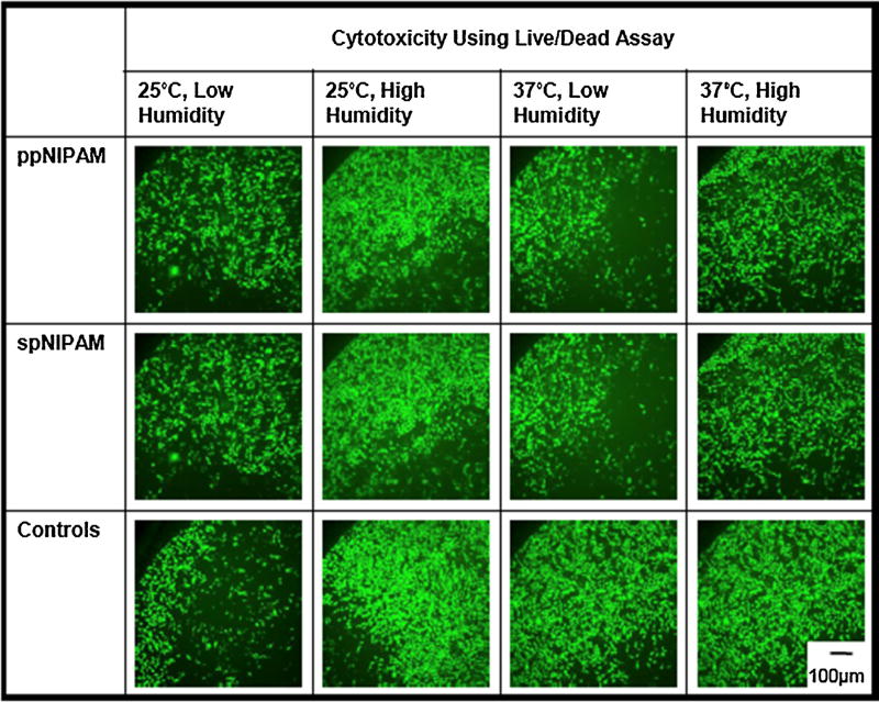
Fig. 4.
Fluorescent microscopy images showing live (green) and dead (red) BAECs after 24 h of incubation with 100% treated media extracted from ppNIPAM (top), spNIPAM (middle), or blank control glass surfaces (bottom). All conditions maintained normal cell growth resulting in live cells after being exposed to treated media. (For interpretation of the references to colour in this figure legend, the reader is referred to the web version of this article.)

