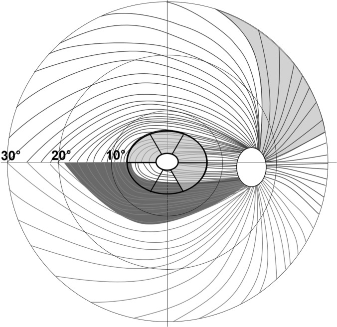
Fig 1. Pattern of the retinal nerve fibre layer (RNFL) and sector map of the macular ganglion cell plus inner plexiform layer (GCIPL) in the right eye.
The central elliptical annulus is divided into six sectors. The light grey areas, RNFL at the 1 and 2 o’clock (superonasal) and GCIPL at the superotemporal, superior, superonasal, and inferonasal sectors were significantly more damaged in non-arteritic anterior ischaemic optic neuropathy (NAION) than in primary open-angle glaucoma (POAG). The dark grey area, RNFL at 7 o’clock (inferotemporal), was significantly more damaged in POAG than in NAION.

