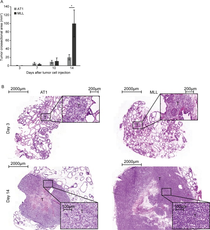
Fig 1. Tumor size.
A) Tumor cross-sectional area at different time points post injection of 2x10⁴ AT1-, or 1x10³ MLL tumor cells into the ventral prostate (bars represent mean +/- SD, n = 7–8 animals/group, * p < 0.05). B) Representative eosin-hematoxylin stained sections showing intraprostatic AT1- and MLL-tumors at day 3 and 14 after tumor cell injection (T; tumor).

