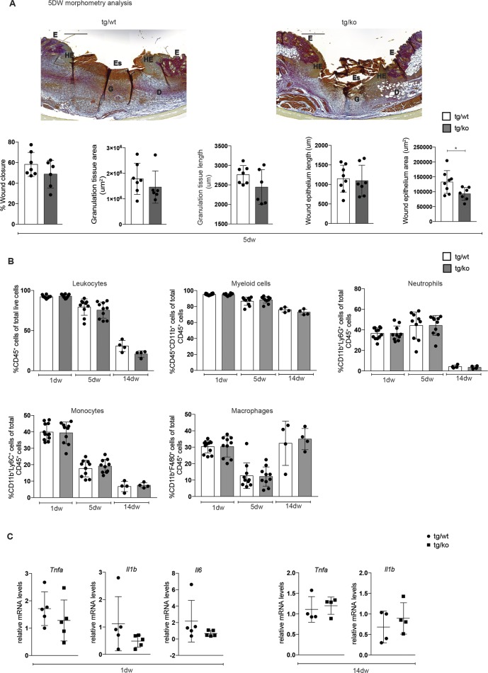
Fig 5. Wound healing is not affected in LysM-Cre Nrf2-ko mice.
(A) Representative pictures of Herovici-stained sections from 5dw of tg/wt and tg/ko mice. Scale bar: 500 μm. D: Dermis; E: Epidermis, Es: Eschar; G: Granulation tissue; HE: Hyperproliferative wound epidermis. Quantitative data from a morphometric analysis of 5dw of tg/wt and tg/ko mice for wound closure, area and length of the granulation tissue, and area and length of wound epithelium are shown below (N = 3–4 mice, n = 6–8 wounds). The raw data of the morphometric analysis of the wounds are shown in S1 Table. (B) Flow cytometry analysis of 1dw, 5dw and 14dw of tg/wt and tg/ko mice for total leukocytes, myeloid cells, neutrophils, monocytes and macrophages. N = 4–6 mice, n = 8–12 wounds. (C) RNA from 1dw and 14dw from control and conditional knockout mice was analyzed for Tnfa, Il1b and Il6 relative to Rps29. Bars indicate mean ±SD. *P ≤0.05; **P ≤0.01; ***P ≤0.001, **** P ≤0.0001 (Mann-Whitney U test).

