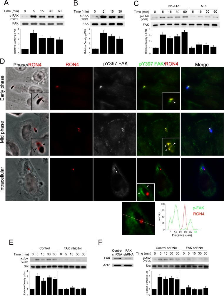
Fig 2. FAK mediates Src activation during T. gondii infection.
A, B, RPE cells (A) or CHO cells (B) were challenged with RH T. gondii for the indicated time points. Cell lysates were used to examine expression of total FAK and phospho-Y397 FAK by immunoblot. C, MIC8KOi parasites were cultured in HFF with or without anhydrotetracycline (ATc; 1 μg/ml). Tachyzoites were harvested and incubated with A549 cells for the indicated time points. Expression of total FAK and phospho-Y397 FAK were assessed by immunoblot. Relative densities of phospho-FAK were compared to that of uninfected (control) cells. Relative density of phospho-FAK for uninfected samples was given a value of 1 Densitometry data represent means ± SEM of 3 independent experiments. D, CHO cells were incubated with YFP T. gondii (RH) for 2.5 min followed by assessment of phospho-Y397 FAK and RON4 expression by immunofluorescence. T. gondii was pseudocolored blue. Arrowheads indicate co-localization of RON4 with phospho-Y397FAK or accumulation of phospho-Y397 FAK around an intracellular tachyzoite. Original magnification X630. Quantification of phospho-Y397 FAK and RON4 signaling intensity for the intracellular parasite is shown underneath. E, A549 cells were incubated with vehicle or FAK inhibitor (1 μM) prior to challenge with RH T. gondii. Cell lysates were obtained and used to probe for total Src and phospho-Y416 Src. A vertical line was inserted between densitometry data of lysates from control and FAK inhibitor-treated cells to indicate that relative densities of phospho-FAK from infected cells treated with or without FAK inhibitor were compared to bands from their respective uninfected (control) cells. Relative density of phospho-FAK for uninfected samples was given a value of 1. F, mHEVc cells transduced with lentiviral vectors that express either FAK shRNA or control shRNA were incubated with RH T. gondii. Immunoblots and densitometries were assessed as above. Experiments shown are representative of 3–4 independent experiments.

