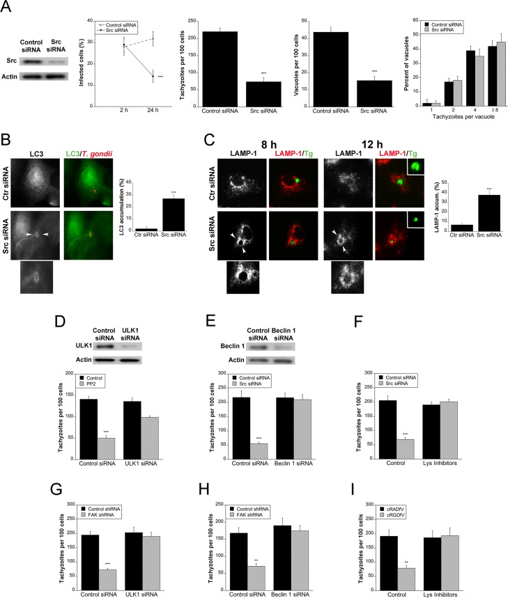
Fig 4. Blockade of Src induces accumulation of the autophagy protein LC3 around the parasite, vacuole-lysosome fusion and killing of the parasite dependent on autophagy the proteins ULK1 and Beclin 1.
A, A549 cells were challenged with RH T. gondii after transfection with either control siRNA or Src siRNA. Monolayers were examined at 2 and 24 h to determine the percentages of infected cells, and at 24 h to ascertain the numbers of T. gondii tachyzoites, T. gondii-containing vacuoles per 100 cells and parasites per vacuole. B, A549 cells transfected with either control siRNA or Src siRNA were challenged with T. gondii-RFP (RH). Expression of LC3 was examined by immunofluorescence 5 h post-challenge. Arrowheads indicate accumulation of LC3 around the parasite. Original magnification X630. C, A549 cells transfected with either control siRNA or Src siRNA were challenged with T. gondii-YFP (RH). Expression of LAMP-1 was examined by immunofluorescence 8 h and 12 h post-challenge. Arrowheads indicate accumulation of LAMP-1 around the parasite. D, mHEVc cells were transfected with control siRNA or ULK1 siRNA followed by treatment with or without PP2 prior to challenge with RH T. gondii. Monolayers were examined by light microscopy 24 h post-infection. E, A549 cells transfected with control siRNA or Src siRNA were transfected with Beclin 1 siRNA. Cells were challenged with RH T. gondii and monolayers were examined by light microscopy 24 h post-infection. F, A549 cells transfected control siRNA or Src siRNA were infected with RH T. gondii. Leupeptin plus pepstatin (Lys Inhibitors) were added post-infection and monolayers were examined microscopically 24 h post-challenge. G-I, mHEVc cells transduced with lentiviral vectors that express either FAK shRNA or control shRNA were transfected with ULK1 siRNA (G), Beclin 1 siRNA (H) or control siRNA followed by incubation with RH T. gondii. mHEVc cells challenged with RH T. gondii were also incubated with leupeptin plus pepstatin (Lys Inhibitors) (I). Monolayers were examined microscopically 24 h post-challenge. Results are shown as the mean ± SEM of 3 independent experiments. ** P < 0.01; *** P < 0.001.

