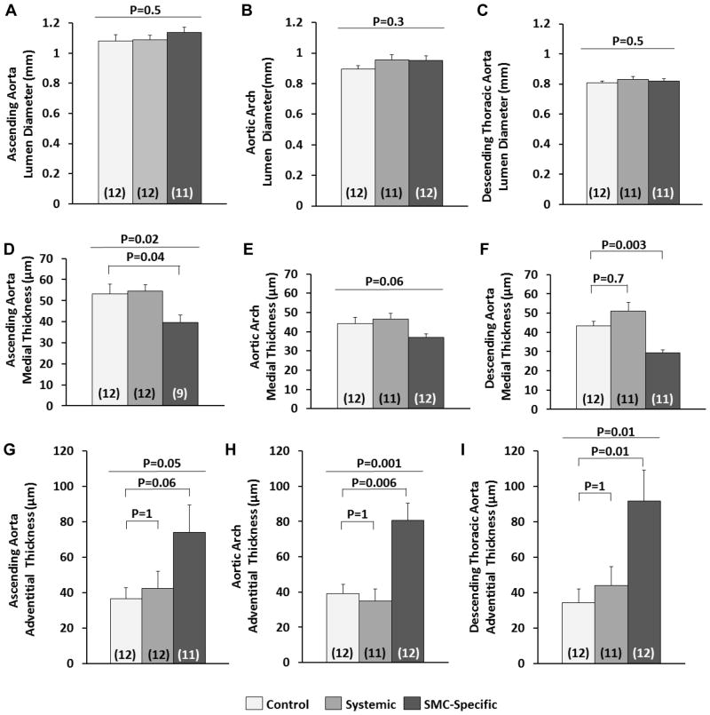
Figure 6.
SMC-specific loss of TGF-β signaling—but not systemic TGF-β signaling blockade—alters thoracic aortic architecture. Planimetry was performed on transverse histologic sections of ascending aorta (A, D, and G), aortic arch (B, E, and H), and descending thoracic aorta (C, F, and I). Aortas were from control mice (no TGF-β inhibition), mice with systemic inhibition of TGF-β activity, and mice with SMC-specific loss of TGF-β signaling. All mice received angiotensin II infusions. Luminal, outer medial, and outer adventitial circumferences were measured. All other parameters were calculated assuming circular geometry in vivo. The number of mice per group is indicated in each bar. Bar heights are means; variance is SEM. (A, B, C–E) P values are from one-way ANOVA (overall P value is above) with Dunnett’s correction for the pairwise comparisons (D). (F–I) P values are from Kruskal-Wallis one-way ANOVA (overall P value is above) with Dunn’s correction for the pair-wise comparisons.

