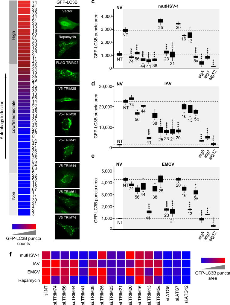
Figure 1. TRIM proteins modulate viral-induced autophagy in a virus species-specific manner.
a, Summary of the TRIM cDNA screen (shown in Supplementary Fig. 1a) in which 61 TRIM proteins were tested for their ability to induce GFP-LC3B puncta formation in transiently transfected HeLa cells. TRIM proteins were categorized into ‘high inducers’ of autophagy (red; defined as inducing more GFP-LC3B puncta than rapamycin stimulation), or ‘low/intermediate inducers’ and ‘non-inducers’ (magenta/blue; defined as inducing fewer GFP-LC3B puncta than rapamycin but more than empty vector transfection, or similar to vector transfection, respectively). b, Representative laser-scanning confocal microscopy images of GFP-LC3B puncta formation in HeLa cells for the top 7 hits from the TRIM cDNA screen shown in (a). Representative images for GFP-LC3B puncta formation induced by rapamycin treatment or empty vector transfection (controls) are also shown. Scale bar, 20 µm. c–e, GFP-LC3B puncta formation in HeLa cells transiently transfected with non-targeting control siRNA (NT) or siRNAs targeting the indicated TRIM proteins and subsequently infected with mutHSV-1 (MOI 4) for 8 h (c), IAV (MOI 5) for 14 h (d), or EMCV (MOI 100) for 6 h (e). Box and whisker plots show the distribution of the area of GFP-LC3B for 3 individual images, each containing ~30 cells. f, Summary of the results of GFP-LC3B puncta formation from the RNAi screen using viral stimuli (c-e) or rapamycin treatment (Supplementary Fig. 1d). NV, no infection. *p<0.05, ***p<0.0005 (ANOVA test). Data are representative of one screen (a), or two (b) or three (c–f) independent experiments.

