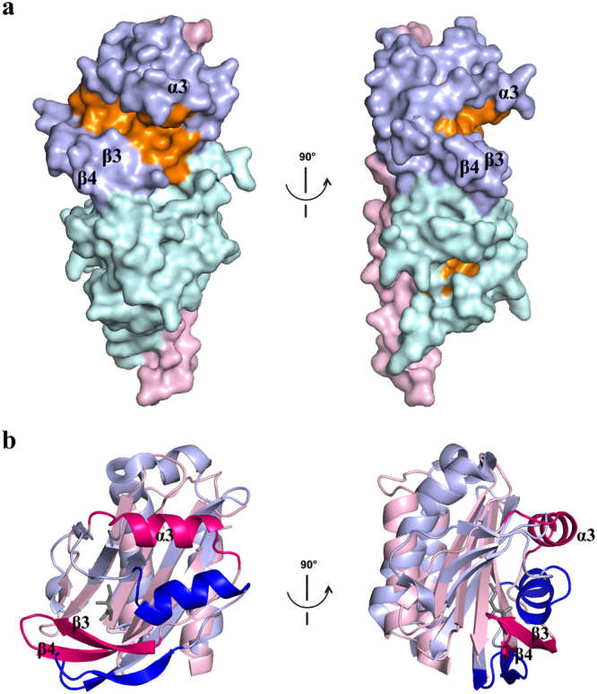
Figure 3.

Putative ligand-binding sites of TlpC LBD. (a) Molecular surface of TlpC LBD with cavities and pockets coloured orange. The stalk helix is coloured pink, the membrane-distal module – light blue and the membrane-proximal module – cyan. (b) Structure superposition of membrane-distal modules of TlpC (pink) and Tlp3 (light blue) highlighting differences in position of helix α3 and β3-β4-tongue (coloured hot pink and blue in TlpC and Tlp3, respectively). Isoleucine bound to the membrane-distal module of Tlp3 is shown as grey sticks.
