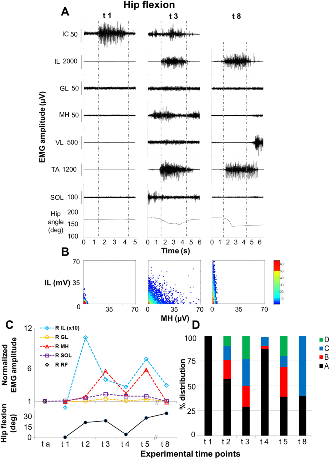
Figure 3.
Volitional attempts to perform hip flexion. Panel (A) Electromyography (EMG) activity and hip joint angle recorded during representative attempts to volitionally perform right hip flexion in the supine position without scES after 80 sessions of locomotor training without scES (t1), after 9.5 months (t3) and after 44 months (t8) of activity-based training with scEs (see Fig. 1 for details). Panel (B) Probability density distribution of EMG amplitudes between the iliopsoas (IL, hip flexor) and the medial hamstrings (MH, hip extensors) calculated during the volitional attempts (data comprised between the two vertical grey dotted lines in Panel A) performed at the experimental time points t1, t3 and t8. Panel (C) EMG amplitude recorded during the volitional attempts, normalized by background (resting) EMG amplitude, and the resulting amount of hip flexion. Kinematics was not recorded at experimental time point t a. Panel (D) Quantitative probability density distribution of EMG amplitudes between iliopsoas and medial hamstrings calculated during the volitional attempts. Black and green indicate the amount of co-contraction at low or high level of activation, respectively (area A and D showed in Fig. 2). Blue and red indicate the isolate activation of iliopsoas or medial hamstrings, respectively (area C and B showed in Fig. 2). EMG was recorded from the following muscles of the right lower limb: IL, iliopsoas; GL, gluteus maximus; MH, medial hamstring; VL, vastus lateralis; TA, tibialis anterior; SOL, soleus. At the experimental time point t a, rectus femoris (RF) was monitored instead of IL as representative hip flexor. EMG activity from intercostal (IC) muscle was recorded to monitor the volitional effort onset.

