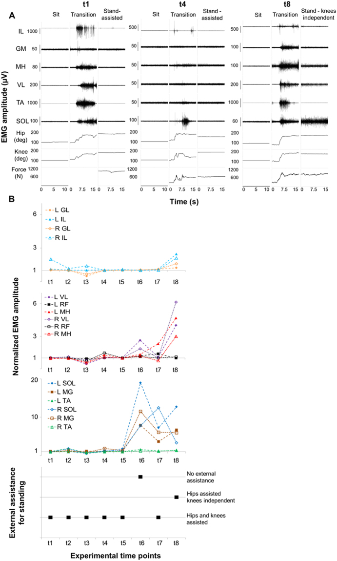
Figure 5.
Time course of EMG amplitude and external assistance during standing. Panel (A) Electromyography (EMG) activity, hip and knee joint angle, and ground reaction forces recorded during sitting, sit-to-stand transition and overground full weight-bearing standing without epidural stimulation prior to any training (t1), after 15.5 months (t4) and after 44 months (t8) of activity-based training with scEs (see Fig. 1 for details). Panel (B) Time course of EMG amplitude recorded during standing, normalized by background (resting) EMG amplitude, and amount of external assistance needed for standing without epidural stimulation. EMG was recorded from the following muscles of the left (L) and right (R) lower limb: IL, iliopsoas; GL, gluteus maximus; MH, medial hamstring; VL, vastus lateralis; RF: rectus femoris; TA, tibialis anterior; SOL, soleus; MG: medial gastrocnemius.

