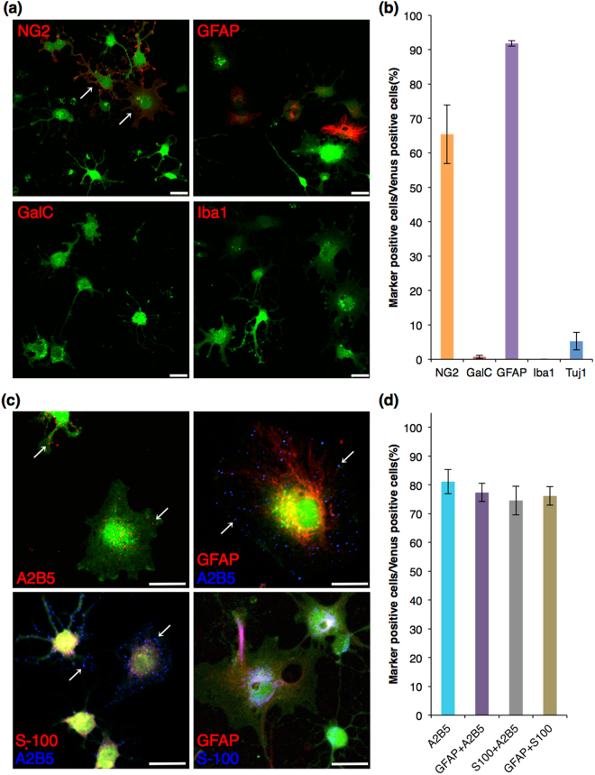
Figure 4.
Immunocytochemistry of glial and neuronal markers in Venus (+) cells after astrocytic differentiation. (a,c) Confocal images of the immunocytochemistry after astrocytic differentiation. Venus fluorescence is shown in green, and each glial marker is shown in red or blue. Venus (+) OPCs developed into flat shaped cells with short primary cell processes (A: arrows). Most of Venus (+) cells expressed A2B5 with GFAP or S-100, or GFAP with S-100. Arrow: punctated staining of A2B5 on Venus (+) cell; Scale bar, 25 µm. (b,d) Cell count analysis showing cell population of Venus (+) cells expressing each marker. Error bars, s.e.m.

