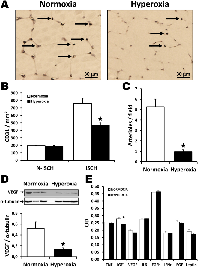
Figure 2.
Effect of neonatal hyperoxia in ischemic tissues. Capillary density (A,B) and arteriolar density (C) in ischemic muscles harvested at day 21 after surgery in the different groups of mice. Arrows in (A) indicate positive (brown) CD31 staining in capillaries. Isch = ischemic. N-Isch = non ischemic. (D) Representative Western blots and quantitative analyses of VEGF expression in ischemic muscles at day 7 after surgery. Data were normalized using loading controls (alpha-tubulin) and are presented as mean ± SEM (n = 4/ group). (E) Effect of hyperoxia exposure on the level of angiogenic factors in the serum, as evaluated by Elisa at day 7 after surgery. *P < 0.05 vs. normoxia.

