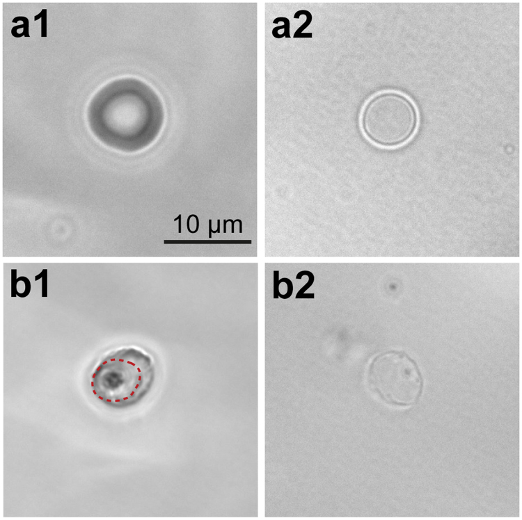
Figure 2.
Optical phase contrast images of uninfected and infected erythrocytes and their ghosts. (a) Uninfected human erythrocytes before (a1) and after (a2) the removal of cytoplasm by osmotic lysis and resealing. (b) The corresponding images of a human erythrocyte infected by P. falciparum (t = 32 h). The parasite and vacuole membrane are visible in an intact erythrocyte (dashed line in b1) but lost after the osmotic lysis and resealing (b2).

