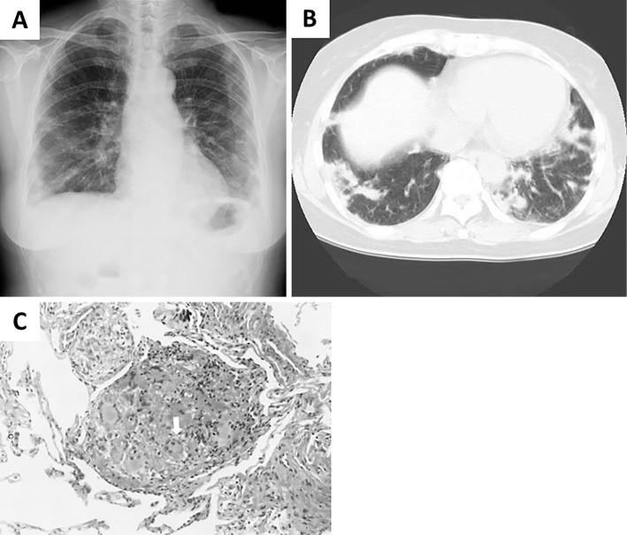
Figure 1.
Diagnostic radiological and histopathological findings related to ILD of unknown etiology. (A) A chest radiograph obtained 10 years ago, showing multiple bilateral nodules. (B) A chest CT scan obtained 10 years ago, showing poorly defined nodules, and peribronchial and subpleural areas of consolidation. (C) Histopathological findings of lung biopsy specimens. Multiple, multinucleated giant cells (white arrow) are observed with inflammatory mononuclear cell infiltration, which is compatible with a granuloma (Hematoxylin and Eosin staining, ×400).

